Chu Hui (Angela) Zeng, 1152626
Dark noise is the noise produced in a photo-detector when the photo-cathode is shielded from all external optical radiation and operating voltages are applied (Desai & Valentino, 2011). Similar to the “snow” on the television screens which is inherent in the design of a video system, dark noise is also inherent in a computed radiography (CR) system. CR employs phosphorous crystals to produce an image. In the imaging plate, with attenuated X-ray photons first converted into light by the crystals, a radiographic image could be created. During the reading process of an image, a certain amount of noise would be unavoidably produced. Therefore, the less the dark noise is, the better the image quality will be.
The dark noise test measures the erasure efficiency of the imaging plate and the CR system (Desai & Valentino, 2011). Thus, the purpose of dark noise test is to assess the level of noise inherent in the system since excessive noise in the plates can compromise image quality (Muhogora, Bonutti, Kazema, Padovani, & Msaki, 2011). In general, this test is recommended to be performed quarterly (Desai & Valentino, 2011).
The procedure for this lab was fairly simple. To perform this test, a 10” × 12” CR cassette was erased first using the Kodak CR reading system. Without any exposure, the cassette was reloaded into the CR reader in order to go through the reading process, and the “Pattern” was set on the CR reader for it. The uniformity of appearance was checked visually and the exposure index (EI) value was recorded. The window width and window level were manipulated in order to visualize the cassette under different viewing conditions of contrast and brightness, which further would show any artifacts the cassette might have. The dark noise test CR lab was conducted in X-ray room 5, B114 of the Institute of Applied Health Sciences on the McMaster University Campus.
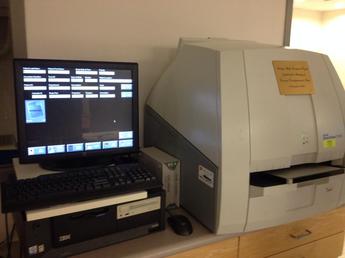 Figure 1. A Kodak Reader in Use to Erase and Read a CR Cassette. | 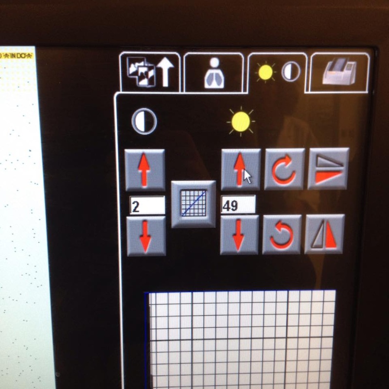 Figure 2. Changing Window Width/ Window Level to Discover Artifacts. |
Qualitative Criteria:
As per American Association of Physicists in Medicine (AAPM) Task Group standards, the qualitative requirement that the dark noise test must meet is that the image produced is uniform without any artifacts, except for collector profile bands in the screen-movement direction. The image obtained from the test is posted below. When closely look at the image, some dots representing dark noise can be clearly seen. By varying the window width and window level, the contrast and brightness of the image was manipulated and no artifacts appeared on it. As a result, the dark noise test passed qualitative criteria set by the AAPM Task Group.
As per American Association of Physicists in Medicine (AAPM) Task Group standards, the qualitative requirement that the dark noise test must meet is that the image produced is uniform without any artifacts, except for collector profile bands in the screen-movement direction. The image obtained from the test is posted below. When closely look at the image, some dots representing dark noise can be clearly seen. By varying the window width and window level, the contrast and brightness of the image was manipulated and no artifacts appeared on it. As a result, the dark noise test passed qualitative criteria set by the AAPM Task Group.
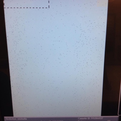
Figure 3. Resultant image from the Dark Noise Test.
Quantitative Criteria:
Other than qualitative criteria, quantitative criteria of the dark noise test is also set by The AAPM Task Group in order to reduce the artifacts that the CR system produces. The guidelines are listed in the figure below, Figure 4.
Other than qualitative criteria, quantitative criteria of the dark noise test is also set by The AAPM Task Group in order to reduce the artifacts that the CR system produces. The guidelines are listed in the figure below, Figure 4.
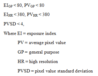
Figure 4. Acceptance Criteria for the Dark Noise Test.
Since a general purpose imaging plate was tested in the lab, an EI value of 80 or less is the acceptance criteria that should be looked for. In this lab, an EI value of 20 was obtained. Since the obtained EI value fell into the standard set by the manufacturer, the CR system in the lab passed the quantitative criteria.
There are some sources of error which can lead to a failure of the dark noise test, such as little damages to the imaging plate. Another example can be that the CR system is not properly quality controlled. If the EI value exceeds the standard demonstrated above, a corrective action needs to be conducted in order to maintain the proper functioning of the CR system, ensuring that the best possible image quality could be provided.
If more than standardized amount of noise is not corrected in time, certain undesired situations could happen. Too much noise on the diagnostic images can either lead to a misdiagnosis of the pathology and this would furthermore impact treatment plan. Even if, fortunately, the misrepresentation is identified right away, a repeat will be requested which would increase the radiation exposure to the patient. Also, with more than needed images being taken, the X-ray suite will age and deteriorate more quickly.
References:
Desai, N., & Valentino, D. J. (2011). A software tool for quality assurance of computed/digital
radiography (CR/DR) systems. Retrieved
from http://www.moderntech.com.hk/sites/default/files/whitepaper/V06_A_software_tool_for_
Quality_Assurance_of_Computed_Digital_Radiography_CR_DR_syste ms.pdf
Muhogora, W., Bonutti, F., Kazema, R., Padovani, R., & Msaki, P. (2011). Performance evaluation of
three computed radiography systems using methods recommended in American Association
of Physicists in Medicine Report 93. Journal of Medical Physics, 36(3), 138-146.
Samei, E., Seibert, J. A., Willis, C. E., Flynn, M. J., Mah, E., & Junck, K. L. (2001). Performance
evaluation of computed radiography systems. Medical Physics, 28(3), 361-371.
 RSS Feed
